X-ray vision: Secrets of the unknown
What do you associate x-rays with? Medical or dental appointments? Long waits at airport security? It may be surprising to know that x-rays are vital in uncovering and examining the interior of objects and specimens in museums. Discover what secrets x-rays have revealed at the Australian Museum!
The Australian Museum is currently undertaking an exciting, decadal-long digitisation project – the Collection Enhancement Project (CEP). The CEP aims to enhance and create better accessibility to the Museum's vast natural science collections, which consist of over 19 million specimens. These millions of specimens have been carefully collected, curated, studied and stored at the AM since it was founded and opened in 1827, making some specimens almost 200 years old! These specimens are a catalogue of our planets biodiversity and are used for scientific research, conservation and education purposes. The CEP employs a number of tools to continue our efforts in future-proofing these priceless collections.
The CEP employs a number of tools to continue our efforts in future-proofing these priceless collections, such as photography and x-ray.
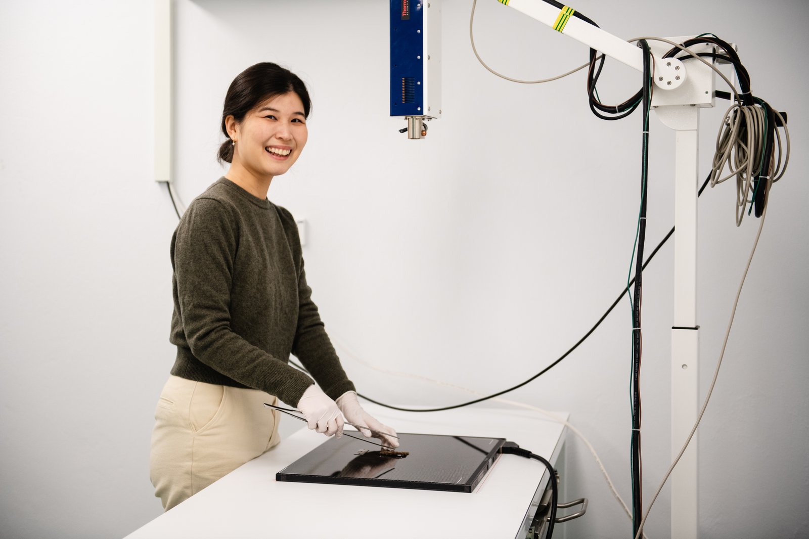
Image: Louise Reily© Australian Museum
Examining X-rays
The use of photography, as a non-invasive way to digitally capture a specimen at a specific point in time, acts to digitally preserve a specimen. Having a digital image further prevents any unnecessary handling that might cause more stress to the specimen, therefore, protecting the continuity of the collection.
However, photography only allows us to view and study the physical exterior of a specimen. A photograph can capture colour, pigmentation, texture, and markings but what if we want to see beyond the visible? The interior bone or skeletal structure? That’s when X-ray vision comes in.
X-ray is another form of digitisation that can be relied upon to reveal an accurate, detailed image of the interior skeletal structure. Specifically for Ichthyology (the study of fishes), x-rays are particularly useful to enable scientists to make accurate counts of the vertebrate and fins (counting spines and rays). They reveal a detailed image of the skeletal structure of the fish, which may assist scientists in determining what species the specimen is or how it relates to other species.
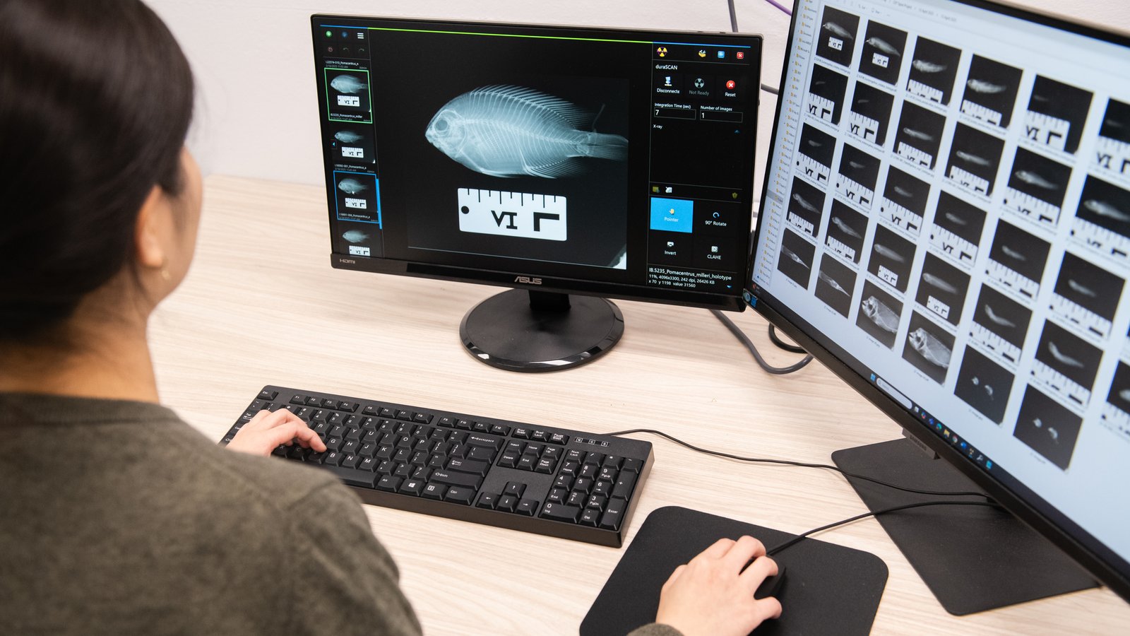
© Australian Museum
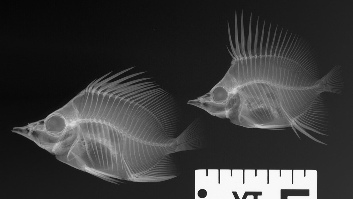
© Australian Museum
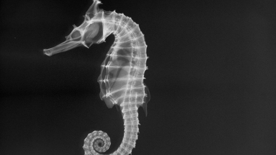
© Australian Museum
The Benefits
- Non-destructive and non-invasive: x-rays do not require any dissecting and cutting of the specimen and allows the specimens to be kept whole for future research
- Accessibility: digital images can be studied by national and international researchers and scientists without loaning specimens, eliminating potential damage of specimens and workload involved in shipping
- Added dimension to scientific research: these scans help scientists describe new species to science, by comparing the interior skeletal structure with the holotype and other specimens
- Quick and cost-efficient tool, compared to other technology
- Unexpected secrets of a specimen, such as their stomach content and diet!
A Secret Revealed
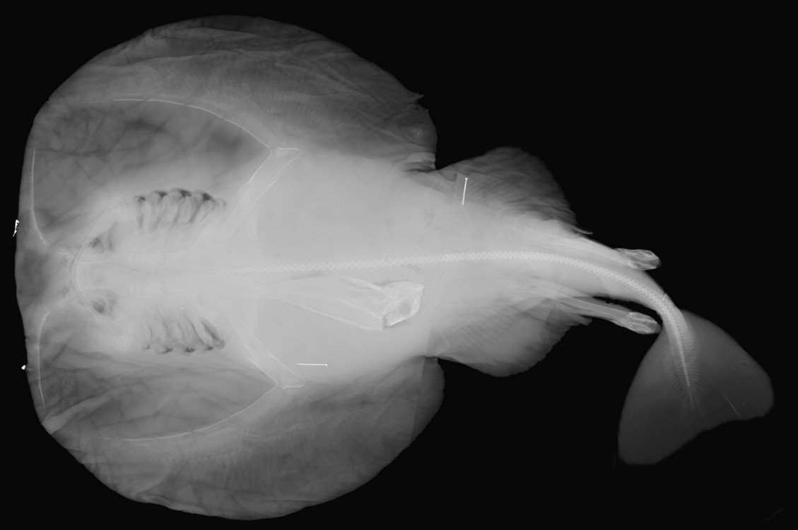
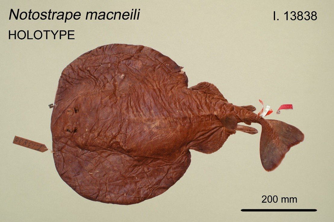
X-rays provide us with multiple benefits and bring us surprises, hidden from the naked eye. For example, a researcher requested an x-ray of a holotype of the Short-tail Torpedo Ray (Tetronarce nobiliana). Upon analysing the x-ray, we discovered it had ingested a whole fish, which was perfectly preserved in the stomach of the Short-tail Torpedo Ray, since 1916 when it was first collected and accessioned into the museum! The stomach specimen was identified as Lepidotrigla, commonly known as Gurnards or Sea Robins from the family Triglidae. Can you spot it in the image below?
© Australian Museum
© Australian Museum
There are currently 15 species belonging to the genus Lepidotrigla. We know from the location where the electric ray was collected, off Green Cape NSW, and that six species of Lepidotrigla inhabit that area. We are unable to determine which one of the six species it is, as identifying the species is very dependent on colour, which cannot be seen. Without the power of x-ray, we would not have even known of the Short-tail Torpedo Ray’s last meal prior to capture and it would still be a mystery. This additional information can be very useful to infer and compare the diet of this fish to its descendants today and whether the changing climate has any effect on its diet.
X-ray is a non-invasive technology that does not require any dissection, preserving the specimen whole for future research and generations to come. It has provided additional important key information such as its diet or the counting of vertebrae to determine a species. The practice of x-raying specimens at the Australian Museum has undoubtedly shifted the way specimens are seen and analysed, from the visible to beyond the visible, which is uncovering hidden secrets yet to be discovered.
Joycelyn Goh, Digitising Technical Officer, Collection Enhancement Project, Australian Museum Research Institute.
Acknowledgement:
I would like to thank Amanda Hay (Ichthyology Collection Manager) and Kerryn Parkinson (Ichthyology Technical Officer) for fact checking and assisting with this blog, as well as Rhiannon Stephens (Life Sciences Digitisation Project Manager) for supporting the publication of the blog.
